Retina is the rear area of the eye where images are captured and sent to the brain, just like a photographic film. Retinal diseases occur when the retina, macula or fovea are affected. Diagnosing retinal diseases is complex process and should be conducted by ophthalmologists with specialization in retinal treatments.
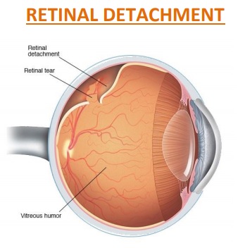
Our eye acts like a camera, it has got lens in the front side and retina in the backside just like reel of camera. (As shown in picture).Lens focuses light and image develops in the retina. If there is no reel in camera then no image can be formed, just like that if there is retinal detachment then image cannot be formed and we lose our eye sight. Retinal detachment is a serious problem; if it doesn’t get treated on time then patient will loss vision and subsequently eye will become small and indrawn.
It depends upon time elapsed after retinal detachment to the date of surgery. After retinal detachment retina becomes weaker day by day. We can attach the retina in its place and vision develops depending on the remaining strength of the retina. So sooner the surgery done more of the vision we can save and more time elapsed after retinal detachment lesser the vision we can save.
If patient get operated within 24 hrs of development of detachment then he is likely to maintain reading and writing vision
If operation is done after 1 week of detachment then he will maintain vision for moving around; fine vision for reading, writing and identifying faces cannot be regained
If surgery is done after one month of detachment then he will maintain only counting finger vision at 1-2 meters
After 6 months of detachment it is difficult to predict how much vision will be preserved.
Small holes and tears are treated with laser surgery or a freeze treatment called cryopexy. These procedures are usually performed in the doctor’s office. During laser surgery tiny burns are made around the hole to “weld” the retina back into place. Cryopexy freezes the area around the hole and helps reattach the retina.
Retinal detachments are treated with surgery that may require the patient to stay in the hospital. In some cases a scleral buckle, a tiny synthetic band, is attached to the outside of the eyeball to gently push the wall of the eye against the detached retina. If necessary, a vitrectomy may also be performed. During a vitrectomy, the doctor makes a tiny incision in the sclera (white of the eye). Next, a small instrument is placed into the eye to remove the vitreous, a gel-like substance that fills the center of the eye and helps the eye maintain a round shape. Gas is often injected to into the eye to replace the vitreous and reattach the retina; the gas pushes the retina back against the wall of the eye. During the healing process, the eye makes fluid that gradually replaces the gas and fills the eye. With all of these procedures, either laser or cryopexy is used to “weld” the retina back in place.
With modern therapy, over 90 percent of those with a retinal detachment can be successfully treated, although sometimes a second treatment is needed. However, the visual outcome is not always predictable. The final visual result may not be known for up to several months following surgery. Even under the best of circumstances, and even after multiple attempts at repair, treatment sometimes fails and vision may eventually be lost. Visual results are best if the retinal detachment is repaired before the macula (the center region of the retina responsible for fine, detailed vision) detaches. That is why it is important to contact an eye care professional immediately if you see a sudden or gradual increase in the number of floaters and/or light flashes, or a dark curtain over the field of vision.